Unit
Compilation 4
Ch. 14 The
digestive System and Nutrition
Table of
Contents
14.1 The digestive system brings nutrients into the
body
a. The walls of the GI tract are
composed of four layers
b. Five basic processes accomplish
digestive system function
c. Two types of motility aid
digestive processes
14.2 The mouth processes food for swallowing
a. Teeth bite and chew food
b. The tongue positions and tastes
food
c. Saliva begins the process of
digestion
14.3 The pharynx and esophagus deliver food to the
stomach
14.4 The stomach stores food, digests protein, and
regulates delivery
a. Gastric juice breaks down
proteins
b. Stomach contractions mix food and
push it forward
14.5 The small intestine digests food and absorbs
nutrients and water
14.6 Accessory organs aid digestion and absorption
a. The pancreas secretes enzymes and
NaHCO3
b. The liver produces bile and
performs many other functions
c. The gallbladder stores bile until
needed
14.7 The large intestine absorbs nutrients and
eliminates wastes
14.8 How nutrients are absorbed
a. Proteins and carbohydrates are
absorbed by active transport
b. Lipids are broken down, then
reassembled
c. Water is absorbed by osmosis
d. Vitamins and mineral follow a
variety of paths
14.9 Endocrine and nervous systems regulate
digestion
a. Regulation depends on volume and
content of food
b. Nutrients are used or stored
until needed
14.10 Nutrition: You are what you eat
a. My Pyramid plan offers a
personalized approach
b. Carbohydrates: A major energy
source
c. Lipids: Essential cell components
and energy sources
d. Complete proteins contain every
amino acid
e. Vitamins are essential for normal
function
f. Minerals: Elements essential for
body processes
g. Fiber benefits the colon
14.11 Weight control: Energy consumed versus energy
spent
a. BMR: Determining how many
Calories we need
b. Energy balance and body weight
c. Physical activity: An efficient
way to use Calories
d. Healthy weight improves overall
health
14.12 Disorders of the digestive system
a. Disorders of the GI tract
1. Lactose intolerance:
Difficulty digesting milk
2. Peptic ulcers: sores
in the stomach
3. Celiac disease
(gluten intolerance)
4. Diverticulitis:
Weakness in the wall of the large intestine
5. Colon polyps:
Noncancerous growths
b. Disorders of the accessory organs
1. Hepatitis:
Inflammation of the liver
2. Gallstones can
obstruct bile flow
c. Malnutrition: Too many or too few
nutrients
d. Obesity: A worldwide epidemic?
14.13 Eating disorders: Anorexia nervosa and bulimia
14.1 The
digestive system brings nutrients into the body

http://classes.midlandstech.com/carterp/Courses/bio211/chap23/Slide2.JPG, Accessed May 3, 2012.
The digestive
system and its accessory organs digest and absorb nearly everything we eat and
drink, regardless of how much we eat or drink (Johnson, 2012). The leftover
non-absorbed waste products and bacteria are stored until they eliminated as
feces. It is a 36-foot journey through the digestive system (The Food Machine).
From table to toilet digestion takes 24 hours (The Food Machine).

http://www.enchantedlearning.com/subjects/anatomy/digestive/color.GIF, Accessed May
3, 2012.
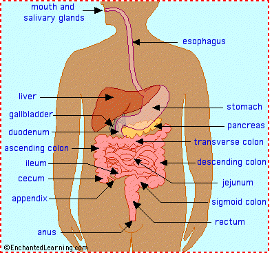
`
The accessory organs of the digestive system include
the (Johnson, 2012):
·
Mouth
·
Pharynx
·
Esophagus
·
Stomach
·
Small
and large intestine
·
Rectum
·
Anus
These organs
form a tube called the gastrointestinal
(GI) tract. The area inside of the tube where food and liquids travel down is
called the lumen.
The digestive system also includes four accessory
organs (Johnson, 2012):
·
The
salivary glands
·
Liver
·
Gallbladder
·
Pancreas
14.1a The walls
of the GI tract are composed of four layers
The walls of the GI tract consist of four layers of
tissue (Johnson, 2012):

http://classes.midlandstech.com/carterp/Courses/bio211/chap23/Slide7.JPG, Accessed May 3, 2012.
1.
Mucosa is the
innermost tissue layer. All nutrients must cross the mucosa to enter the blood.
2.
Submucosa is the middle
layer of connective tissue containing blood vessels, lymph vessels and nerves.
Components of food that are absorbed across the mucosa enter the blood and
lymph vessels of the submucosa.
3.
Muscularis is the layer
that is responsible for movement. The muscularis consist of two or three sub
layers of smooth muscle.
4.
Serosa is the
outermost layer and is a thin connective tissue that surrounds and protects the
other three layers and attaches the digestive system to the walls of the body
cavities.
Sphincters are thick rings of circular smooth
muscle that separates some of the organs of the GI tract from each other.
14.1b Five basic
processes accomplish digestive system function
The digestive
system receives food, takes it apart so that the nutrients can be absorbed into
the body. Food is the starting point of the digestion process (The Food Machine).
Digesting food takes 10% of our energy required, 70% expended in keeping the
body idling, running the processes that keep us alive, and 20% is left for our
energy output for all our activities during the day (The Food Machine).
This involves five basic processes (Johnson, 2012):
1.
Mechanical
processing and movement is accomplished by chewing. Chewing breaks food into
smaller pieces, and two types of movement mix the contents of the lumen and
propel the pieces forward
2.
Secretion is fluid that
consists of digestive enzymes, acid, alkali; bile and mucus is secreted into
the GI tract at various places. Several hormones regulate digestion are
secreted into the bloodstream as well.
3.
Digesting the contents of
the lumen is broken down both mechanically and chemically into smaller and
smaller particles, culmination in nutrient molecules.
4.
Absorbing nutrient
molecules as they pass across the mucosal layer of the GI tract and into the
blood or lymph
5.
Eliminating undigested
material from the body by the anus
14.1c Two types
of motility aid digestive processes
The smooth muscles of the GI tract produces two
kinds of motility (movement) called (Johnson, 2012):
1.
Peristalsis is the movement
that propels food forward by causing the smooth muscles in the GI tract to
contract. This peristaltic wave of contraction ripples through the organs of
the GI tract, mixing the contents of the stomach and pushing the contents of
the esophagus and intestines forward. Peristalsis occurs in all parts of the GI
tract, but is most prevalent in the esophagus,
where it transports food rapidly to the stomach.
2.
Segmentation mixes food by
causing the smooth muscles in the GI tract to contract and relax in random
fashion. This results in the sloshing movement of the contents in the lumen
mixing the food and liquid together. Food particles are then pressed against
the mucosa, enabling the body to absorb their nutrients. Segmentation occurs
primarily in the small intestine, as food is digested and absorbed.
14.2 The mouth
processes food for swallowing
The mouth is the
entrance to the GI tract where digestion begins with the process of chewing,
breaking food into smaller particles.
14.2a Teeth bite
and chew food
The teeth chew
food into smaller pieces enabling us to swallow.
There are four types of teeth created for different
purposes (Johnson, 2012):


http://classes.midlandstech.com/carterp/Courses/bio211/chap23/Slide11.JPG, Accessed May
3, 2012.
1.
Incisors have a sharp-edge
and cut food.
2.
Canines are pointed and
tear food
3.
Premolars grind and crush
food
4.
Molars grind and crush
food as well
Children have 20
teeth that they develop by age two and gradually replace by permanent teeth
until they reach the full number of adult teeth. Most adults have 32 permanent
teeth (Johnson, 2012).
Each tooth sits
in a socket in the jawbone lined with periodontal membrane. Each tooth consist
of a crown (the outside portion of
the tooth you can see) which is covered by a layer of enamel, which can repair minor damage to its outer part (The Food
Machine) and is an extremely hard nonliving compound of calcium and phosphate.
Beneath the enamel is dentin (a
bonelike living layer). The soft innermost pulp cavity contains the blood
vessels that supply the dentin, as well as the nerves that cause pain (Johnson,
2012). The region below the gum line is the root.
Bacteria that
remain inbetween our teeth cause cavities.
The bacteria release acids that can dissolve enamel, creating cavities. If not
treated, cavities can deepen, eroding the dentin and pulp cavity causing a toothache.
Tooth decay may inflame the soft gum tissue around the tooth, causing
gingivitis. Periodontitis is decay
that inflames the periodontal membrane. Regular brushing and flossing will
ensure good dental hygiene.
14.2b The tongue
positions and tastes food

http://classes.midlandstech.com/carterp/Courses/bio211/chap23/Slide9.JPG, Accessed May 3, 2012.
The tongue is a skeletal muscle that
positions food over the teeth and mashes it against the roof of the mouth. The
tongue is an essential element in tasting food and for talking.
14.2c Saliva
begins the process of digestion
Saliva moistens food, making it easier to chew
and swallow. The thought of food is enough to make your mouth water. Three
pairs of salutatory glands produce two pints of saliva every day. The saliva
pours through miniature fountains from under your tongue (video).
Saliva contains four main ingredients (Johnson,
2012):
1.
Mucin is a mucus-like
protein that holds food particles together so they can be swallowed more
easily.
2.
Slivary amylase is an
enzyme that begins the process of digesting carbohydrates.
3.
Bicarbonate in salvia maintains
the pH of the mouth between 6.5 and 7.5, the range over which salivary amylase
is most effective.
4.
Lysozyme inhibits
bacterial growth.
14.3 The pharynx
and esophagus deliver food to the stomach
The stomach stores ingested food and water
until it is delivered to the small intestine. It also secretes a strong acid
(HCI) hydrochloric acid that breaks
down proteins and kills bacterial.
The stomach is
covered with deep pits lined with microscopic cells that release HCI acid while
their neighbors secrete a sticky mucous that coats the stomach (The Food
Machine). This protects the stomach from approximately a gallon of gastric
juices the stomach lining pours into the stomach (The Food Machine).
After we have
chewed our food and mixed it with saliva the tongue pushes it into the pharynx (throat) for swallowing.
Swallowing involves a sequence of events that is
coordinated with a temporary halt in breathing (Johnson, 2012):
1.
Swallowing begins as the
tongue pushes a lump of food (bolus) into the pharynx.
2.
Before
swallowing, muscles keep the esophagus closed. The presence of food stimulates
receptors in the throat and initiates the swallowing
reflex.
3.
The
soft palate rises to close off the
passageway into the nasal cavity and the larynx rises slightly.
4.
The
epiglottis bends to close off the
airway to the trachea temporarily and opens the esophagus.
After the throat,
is the esophagus a muscular tube
consisting of both skeletal and smooth muscle that connects the pharynx to the
stomach. The lining of the esophagus produces mucus that helps food slide
easily.

http://classes.midlandstech.com/carterp/Courses/bio211/chap23/Slide14.JPG, Accessed May
3, 2012.
Acid reflux or heartburn is caused by the sphincter
malfunctioning. The sphincter prevents reflux of the stomach’s contents back
into the esophagus. Prolonged heartburn may cause esophageal ulcers because
stomach acid can erode the mucosa of the esophagus.
14.4 The stomach
stores food, digests protein and regulates delivery
The stomach is a muscular, expandable sac.

http://classes.midlandstech.com/carterp/Courses/bio211/chap23/Slide15.JPG, Accessed May
3, 2012.
The stomach performs three important functions
(Johnson, 2012):
1.
Stores
food until it is digested and absorbed. The stomach can expand 1-3 liters of
capacity when we eat.
2.
The
stomach digests proteins using strong acid and protein-digesting enzymes. The
acid also kills most bacteria. Muscle contractions mix and break apart the food
particles and push the mixture into the small intestine.
3.
Regulates
the rate at which food is delivered into the small intestine.
14.4a Gastric
juice breaks down proteins
The walls of the stomach consist of four layers the
same as the GI tract (Johnson, 2012):

http://classes.midlandstech.com/carterp/Courses/bio211/chap23/Slide17.JPG, Accessed May
3, 2012.
1.
Mucosa
2.
Submucosa
3.
Muscularis
4.
Serosa
Gastric juices
consist of hydrochloric acid (HCI) (some of the cells lining the glands secrete
(HCI) and pepsinogen (a large
precursor molecule that becomes a protein-digesting enzyme called pepsin once
exposed to stomach acid). The pepsin and acid dissolve the connective tissue in
food and digest proteins and peptides into amino acids so they can be absorbed
in the small intestine.
The stomach
produces 1-2 liters gastric juices immediately after meals. Chyme is the watery mixtures of
partially digested food and gastric juice that is delivered to the small
intestine. The pyloric sphincter
between the stomach and the small intestine regulates the rate of transport of
chime into the small intestine.
Gastric juices
to not digest in the stomach because some of the cells lining the stomach and
the gastric glands continuously produce a protective barrier of mucus. A peptic ulcer is an open sore that
develops when gastric juices become in contact with the living cells instead of
the mucus.
14.4b Stomach
contractions mix food and push it forward
When you eat,
your stomach does not contract and allows it to relax and stretch. Stretching
signals peristalsis to increases. While your stomach is empty, muscle
contractions keep it small.
Peristalsis pushes the chyme toward the pyloric
sphincter in a forward and backward movement, squeezing it, causing it to mix.
Each contraction propels about a tablespoon of chyme into the small intestine
before the pyloric sphincter closes.
It takes two to
six hours for the stomach to empty completely after a meal (Johnson, 2012).
Peristalsis is more forceful when the stomach is full then when empty. Chyme
with a high acid or fat content stimulates the release of hormones that slows
stomach peristalsis, giving the small intestine more time to absorb the
nutrients. The stomach does not absorb nutrients because it lacks cellular
transporting mechanisms and because its inner lining is coated with mucus.
14.5 The small
intestine digests food and absorbs nutrients and water
Nutrients and
water are absorbed in the small intestine. If our small intestines were not
neatly wrapped insides of us, we would have to be 30ft tall (The Food Machine).
Enzymes from the pancreas and the small intestine break down carbohydrates and
fats so that they can be absorbed. The inner surface of the small intestine has
many villi and microvilli, which
increase the surface area of absorption. This surface area is ten times the
area of our skin, enough to carpet a living room (The Food Machine).

http://classes.midlandstech.com/carterp/Courses/bio211/chap23/Slide30.JPG, Accessed May
3, 2012.
The small intestine has two major functions (Johnson,
2012):
1.
The
stomach partially digests proteins to smaller peptides, under the influence of
strong acids and pepsin. Protein digestion continues in the small intestine,
but also digests carbohydrates and lipids, which involve neutralizing the
highly acidic gastric juice and adding additional digestive enzymes from the
intestine and pancreas.
2.
Eventually
the proteins, carbohydrates, and lipids in food are broken down to single amino
acids, monosaccharides, fatty acids and glycerol, which are small enough to be
transported across mucosal cells into the blood. Nearly 90% of the absorbable
nutrients and water is absorbed in the small intestine.
The small intestine consists of three different
regions (Johnson, 2012):
1.
Duodenum is where most
of the digestion takes place.
2.
Jejunum is where the
rest of the products of digestion are absorbed.
3.
Ileum is also where
the rest of the products of digestion are absorbed.
The structure of
the small intestine contains villi attached to the mucosa. Each epithelial cell of the villi has dozens
of even smaller, cytoplasmic projections called microvilli, which gives the mucosal surface a velvety appearance.
Combined the folds of villi and microvilli enlarge the surface area of the
small intestine increasing its ability to absorb nutrients. At the center of
each villus are capillaries and a small lymph vessel called a lacteal to transport nutrients to
larger blood vessels and lymph vessels.
14.6 Accessory
organs aid digestion and absorption
The digestive system has four accessory organs
(Johnson, 2012):
·
Salivary
glands
·
Pancreas
·
Gallbladder
·
Liver
14.6a The
pancreas secretes enzymes and NaHCO3
The pancreas is an elongated organ that
lies just behind the stomach and has both endocrine
(secretes hormones that regulate blood glucose levels) and exocrine functions.

http://classes.midlandstech.com/carterp/Courses/bio211/chap23/Slide38.JPG, Accessed May
3, 2012.
The pancreas produces and secretes the following
(Johnson, 2012):
·
Digestive
enzymes include proteases (enzyme
that digest proteins) and lipase a
lipid-digesting enzyme.
·
Sodium
bicarbonate
(NaHCO2) functions to neutralize stomach acid.
14.6b The liver
produces bile and performs many other functions
The liver is a large organ located in the
upper right abdomen cavity with 500 different functions (The Food Machine).
The livers
primary digestive function is to facilitate the digestion and absorption of
lipids by producing bile. Bile is a
watery mixture containing electrolytes, cholesterol, bile salts derived from
cholesterol, a phospholipix called lecithin,
and pigments (primarily bilirubin) derived from the breakdown of hemoglobin.
The bile salts break lipids down
into smaller droplets that are digested by lipases
(lipid-digesting enzymes) from the pancreas.
The hepatic portal system in the digestive
system carries nutrient-rich blood directly from the digestive organs to the
liver by the hepatic portal vein.
The liver begins processing and storing nutrients for the body just as soon as
digestion and absorption has begun. After passing through the liver, the blood
is returned to the general circulation.

http://classes.midlandstech.com/carterp/Courses/bio211/chap23/Slide32.JPG, Accessed May
3, 2012.
The liver severs other functions that maintain
homeostasis (Johnson, 2012):
·
Stores
fat-soluble vitamins (A, D, E and K) and iron
·
Stores
glucose as glycogen after a meal and converts glycogen to glucose between meals
·
Manufactures
plasma proteins like albumin and fibrinogen from amino acids
·
Creates
and stores some lipids
·
Inactivates
many chemicals like alcohol, hormones, drugs and poisons
·
Converts
ammonia (NH3), a toxic waste product of metabolism into less toxic urea
·
Destroys
worn-our RBCs.
Overexposure to
toxic chemicals, medications or alcohol can damage the liver because it takes
up these substances to “detoxify” them, killing some liver cells in the
process. Long-term exposure to any of the above-mentioned toxins can destroy
enough cells to impair liver functions, known as cirrhosis.
14.6c The
gallbladder stores bile until needed

http://classes.midlandstech.com/carterp/Courses/bio211/chap23/Slide27.JPG, Accessed May
3, 2012.
The bile
produced by the liver flows through ducts to the gallbladder, which concentrates bile by removing most of the water,
and stores it until after a meal. Then the bile is secreted into the small
intestine by the bile duct, which joins the pancreatic duct.
14.7 The large
intestine absorbs nutrients and eliminates wastes
By the time the
contents of the digestive tract reach the large intestine, most of the
nutrients and water have been absorbed. The large intestine absorbs most of the remaining nutrients and water
and stores the now nearly solid waste material until it is eliminated. The
large intestine is larger in diameter than the small intestine but is shorter.
It begins as a pouch called the cecum,
which receives the chyme from the small intestine. The fingerlike appendix
extends from the cecum. The appendix
has no known digestive function, but is known to become inflamed and can be
removed (Johnson, 2012).
Most of
the large intestine consists of four regions called the colon (Johnson, 2012):

http://classes.midlandstech.com/carterp/Courses/bio211/chap23/Slide40.JPG, Accessed May 3, 2012.
1.
The
ascending colon rises along the
right side of the body.
2.
The
transverse colon crosses over to the
left side
3.
The
descending colon passes down the
left side to the last colon
4.
Sigmoid colon is where feces are
stored until defecation where they
pass through the rectum to the anus.
Feces are indigestible material that contains
some bacterial. Some of these bacteria release by-products that are useful to
us like vitamin K (important for blood clotting). Some bacteria also produce
less helpful substances such as intestinal
gas, a by-product of metabolism as they break down food.
A neural reflex controls defecation. The
internal anal sphincter consists of a ring of smooth muscle that normally keeps
the anus closed (Johnson, 2012). When feces enter the rectum, the rectum
becomes stretched and the neural reflex causes the internal anal sphincter to
relax and the rectum to contract, expelling the feces. We have control over the
external sphincter by voluntarily contracting this muscle, which allows us to
hold our pee or poop until we want to expel these bodily fluids.
14.8 How
nutrients are absorbed
Your body
absorbs nutrients differently depending on the type of nutrient.
14.8a Proteins
and carbohydrates are absorbed by active transport

http://classes.midlandstech.com/carterp/Courses/bio211/chap23/Slide48.JPG, Accessed May 3, 2012.
In the small
intestine, enzymes from the pancreas and enzymes secreted by the mucosal layer
of the stomach and from the small intestine break down proteins into amino
acids. The amino acids are actively transported into the mucosal cells and
eventually move by facilitated diffusion out and make their way to the
capillaries.
Carbohydrate
digestion begins in the mouth, where salivary
amylase breaks down polysaccharides into disaccharides. It is completed in
the small intestine with the addition of pancreatic amylase and enzymes from
the small intestine. Together these enzymes break down the remaining
carbohydrates into monosaccharide’s
(simple sugars such as glucose) and are then actively transported by proteins.
14.8b Lipids are
broken down, then reassembled

http://classes.midlandstech.com/carterp/Courses/bio211/chap23/Slide49.JPG, Accessed May 3, 2012.
Bile salts emulsify lipids into small fat droplets
and are digested by pancreatic and intestinal lipases. The product is monoglycerides,
which dissolve in micelles. Micelles
are small droplets composed of bile salts and lecithin that transport fatty acids and monoglycerides to the outer
surface of the mucosal cells so that they can be absorbed into the cells. Once
inside the cells, the fatty acids and monoglycerides recombine into triglycerides,
coated with proteins to form water-soluble droplets called chylomicrons. They are released from the cell by exocytosis. They are too large to enter
capillaries so they travel in the lymph vessels until the lymph returns to the
venous blood vessels near the heart.
14.8c Water is
absorbed by osmosis
A high
concentration of water in the lumen represents a strong driving force for the diffusion
of water through the epithelial layer of the cells of the small intestine and
into the blood (Johnson, 2012). Diarrhea
is caused when the small intestine delivers too much food residue to the large
intestine. Constipation is caused when
feces remain in the large intestine and colon so long that too much water is
absorbed. The feces become dry and hard making it hard to poop. Water makes up
60% (10 gallons) of the human body and is constantly being reclaimed by the
body through our waste products (The Food Machine). The lining of the digestive
tract changes as we enter into the large intestine from cells that are designed
to absorb nutrients to cells that are designed to filter out water (The Food
Machine). Digestive juices turn food into slush and the body extracts two
thirds of the water out by the large intestine (The Food Machine). The
intestine walls soak up ten pints of water every day and a network of
capillaries carry the absorbed water and nutrients away from the intestines and
back into circulation (The Food Machine).
14.8d Vitamins
and minerals follow a variety of paths.
Vitamins are
absorbed differently depending on if they are fat-soluble or water-soluble. Fat-soluble vitamins dissolve in the
micelles and are absorbed by diffusion across the lipid membrane of the mucosal
cell layer. Water-soluble vitamins
are absorbed by either active transport or diffusion through channels or pores.
Minerals (ions) like, sodium,
potassium, calcium, phosphate, sulfate and magnesium are electrically charged
and not lipid soluble (fat-soluble).
The body digests
and reabsorbs the components of the digestive secretions themselves. Water and
minerals in the digestive secretions are reabsorbed by the normal mechanisms
for these nutrients. Enzymes are digested to their component amino acids and
the amino acids are then reabsorbed. Bile salts are reabsorbed, returned to the
liver and used again.
14.9 Endocrine
and nervous systems regulate digestion
Regulation of
the digestive system involves altering the movement of secretions of various
organs so that each operates efficiently. The digestive process alters the
internal environment temporarily because of the absorbed nutrients enter the
blood in a short time. This is different from most regulatory mechanisms that
operate to maintain homeostasis.
14.9a Regulation
depends on volume and content of food
The endocrine system and nervous system regulate digestion
according to both the volume and content of food. Most digestion and absorption
occurs in the stomach and small intestine. When the stomach stretches, neural
reflexes increase stomach peristalsis and secretion of gastric juices. This
releases the hormone gastrin, which triggers
the release of more gastric juice.
When chyme enters the small intestine, the stretching
of the duodenum increases segmentation to mix the chyme.
The duodenum secretes two hormones into the
bloodstream (Johnson, 2012):
·
Secretin
·
Cholecystokinin
The acid in chyme releases secretin, which stimulates the pancreas
to secrete water and bicarbonate to neutralize acid. Fat and protein stimulate
release of cholecystokinin (CCK)
which signals the pancreas to secrete enzymes that aid indigestion.
If chyme flows
too quickly from the stomach, the small intestine will slow stomach activity.
Gastrin and a neural reflex involving stretching of the stomach increase
movement of the large intestine after eating.
14.9b Nutrients
are used or stored until needed
All cells need nutrients, which are substances in food
that are required for growth, reproduction and the maintenance of health.
Nutrients in your last meal are used to fuel cellular activities, build cell
components and serve other vital functions. Your cells draw these nutrients
from your blood and your blood obtains them from your digestive system or from
nutrient storage pools (Johnson, 2012).
Once nutrients
are absorbed, they must be used whether they are consumed immediately, stored
until later or combined with more nutrients to create other molecules.
14.10 Nutrition:
You are what you eat
It matters what
you eat. Poor nutrition is associated with diseases ranging from cancer to cavities.
Good nutrition, on the other hand, improves overall health and lowers the risk
of health problems.
14.10a My
Pyramid plan offers a personalized approach

http://healthvermont.gov/prevent/diabetes/mypyramid.gif, Accessed May 3, 2012.
My Pyramid is the recommended dietary guidelines
that include physical activity as well as healthy nutrition. The pyramid
indicates the recommended consumption of foods from six different groups and
gives you recommendations on what to eat from each group.
My Pyramid is
highly interactive. If you enter your age, gender and activity level on the Web
site, the system will match you with the best plan you need bases on how the
required caloric intake (Johnson, 2012). The feature, My Pyramid Tracker, lets
you compare your current eating and physical activity patterns to
recommendations in the Dietary Guidelines and track them for up to a year to
follow your progress.
My Pyramid has
come under controversy with some nutritionists for instance: My Pyramid
recommends that to prevent osteoporosis, adults should drink three glasses of
low fat milk per day. That is over 300 calories just in milk. This is not
necessary for everyone. Nevertheless, My Pyramid is a good place to start for
the basics.
General recommendations for a healthy diet include
(Johnson, 2012):
·
Eat
a variety of foods
·
Maintain
a healthy weight
·
Eat
plenty of fruits, vegetables and whole-grain products
·
Choose
a diet low in cholesterol and saturated fat
·
Use
sugar in moderation
·
Consume
salt and sodium in moderation (1tsp of salt per day)
·
Drink
alcohol in moderation.
14.10b
Carbohydrates: A major energy source
Carbohydrates are one of the body’s main sources of
energy and many nutritionists recommend that approximately 45-65% of our
Calorie intake come from carbohydrates. A
calorie is a measure of energy.
Carbohydrates
may be either simple or complex (Johnson, 2012). Simple carbohydrates (sugars) are found in natural foods such as
fruit and honey. Complex carbohydrates
such as starch or glycogen consist of many sugar units linked together. Whole
foods containing complex carbohydrates are better for us than refined sugars (corn sweeteners,
dextrose or fructose) because they release sugars more slowly and contribute fiber,
vitamins, and minerals. In the body, stored starch and glycogen are broken down
to glucose, one of the premier sources of energy.
14.10c Lipids:
Essential cell components and energy sources
Lipids are essential components of every
living cell. Phospholipids and cholesterol make up most of the cell membrane. Cholesterol also forms the backbone of
steroid hormones and is used to make bile. Fat stores energy, cushions organs,
insulates the body and stores several vitamins. Most of the fats in food are
triglycerides, which consist of three fatty acids attached to a glycerol
molecule.
Fats fall into two categories (Johnson, 2012):
·
Saturated fats are solid at
room temperature, found primarily in meat and dairy products and in a few plant
sources such as coconut, and palm kernel oil. They raise blood levels and are
associated with atherosclerosis and heart disease.
·
Unsaturated fats
are liquids (oils)
at room temperature and considered healthier than saturated fats because they
tend to lower cholesterol levels. Unsaturated fats found in olive, canola, safflower,
and corn oils and derived from plants. Certain cold-water fish (salmon, trout
and sardines) are rich in omega-3 fatty acids, polyunsaturated fatty acids that
are linked to reduce risk of heart disease.
Tran’s fats made from unsaturated fats. Unsaturated
fats are missing one or more pairs of hydrogen atoms in their fatty acid tails
by partial hydrogenation reconfigures the positions of some of the remaining
unpaired hydrogen atoms trans fat is created. Tran’s fat can be found in
deep-frying oil, baked goods (cookies, crackers, snacks) and in vegetable
shortening and margarine. Tran’s fats tend to raise cholesterol and increase
the risk of cardiovascular heart disease.
The liver can
make cholesterol and most of the lipids the body needs, but cannot create the
essential fatty acids. These fatty acids like linoleic and linolenic acids
(present in corn and olives) must be consumed as food. Linoleic and linolenic
acids are important for proper cell membrane structure and can be obtained by
eating a tsp of corn or olive oil a day to satisfy the daily requirements.
We must be
careful of how many lipids we consume. Diets high in saturated fat, cholesterol
and Trans fats increase our risk for developing cardiovascular disease and
certain cancers.
14.10d Complete
proteins contain every amino acid
Proteins make up the enzymes that direct metabolism,
serve as receptor and transport molecules and build our muscle fibers. They are
vital components of every cell. All proteins are composed of 20 different amino
acids. The body can make 12 of these amino acids; the other eight that the body
cannot produce is called the essential
amino acids. The body must ingest these eight amino acids (isoleucine,
leucine, lysine, methionine, phenylalanine, threonine, tryptophan, and valine)
(Johnson, 2012). A complete protein contains all 20 amino acids in proportions
that meet our nutritional needs. Foods like trail mix, nuts, soybeans, hummus,
red beans and rice will provide the necessary essential amino acids.
Every enzyme has
a unique amino acid sequence, even if one amino acid is missing in the diet,
the body may be unable to produce enzymes the proper time in development.
Protein deficiencies during pregnancy and childhood can retard growth and alter
physical and mental performance.
14.10e Vitamins
are essential for normal function
Our bodies can
produce only a few vitamins; our skin creates vitamin D when we are exposed to
sunlight, and bacteria living in the colon manufacture vitamins K, B6 and
biotin. All other vitamins must be ingested.
Vitamins fall into two categories (Johnson, 2012):
·
Fat
soluble
·
Water soluble
The difference
between the two is how they are absorbed and stored. Fat-soluble vitamins are absorbed more readily if there is fat in
the diet and are stored in fat tissue and releases as needed. Water-soluble vitamins are stored
briefly and rapidly excreted in urine. Consuming water-soluble vitamins on a
regular basis is necessary.
14.10f Mineral:
Elements essential for body processes
Minerals are the ions in blood plasma and cell cytoplasm (sodium, potassium, chloride,
and many others). They represent most of the chemical structure of bone
(calcium, phosphorus and oxygen). They also contribute to the activity of
nerves and muscles (sodium, potassium and calcium).
The National
Research Council publishes the current best estimate of how much vitamins and
minerals we need daily called the (RDA) the Recommended Dietary Allowance
(Johnson, 2012). If you eat a balanced diet of whole foods you can achieve t he
RDA without taking supplements.
14.10g Fiber
benefits the colon
Fiber is found in many vegetables, fruits and
grains (indigestible). Even though our bodies cannot digest it, it is
necessary. Fiber makes feces bulky and helps them pass easily through the
colon.
A low-fiber diet
can lead to constipation, hemorrhoids
(swollen veins in the lining of the anus, caused by straining when pooping) and
a disorder called diverticulitis. Acquiring not enough fiber has been
associated with and increased risk of developing colon cancer.
14.11 Weight
control: Energy consumed versus energy spent
To maintain a
constant body weight, energy intake must equal energy expenditure. Any consistent
imbalance in energy intake versus energy expenditure over time leads to weight
gain or loss. Exercise can dramatically change how many calories we expend. To
lose weight we must eat less, exercise more, or do a little of both. ScienceDaily
2012 states in their article that counting calories may be only part of the
weight loss equation. According to scientists, sleep is a major requirement for
weight loss. They did a study on mice to show how the “rev-Erb alpha” can lead
to excessive weight gain and related health problems. The rev-Erb alpha is our
internal body clocks. This experiment provides new insights into the importance
of proper alignment between the body’s internal timing and natural
environmental light cycles to prevent or limit excessive weight gain and the
problems this weight gain can cause (ScienceDaily, 2012).
The body
requires energy to fuel metabolic processes and other activities. When we
digest nutrients, energy is obtained.
Energy is
measured in units called calories. A calorie
is the amount of energy needed to raise the temperature of 1 gram of water by 1
degree Celsius (Johnson, 2012). In Biology, a calorie (1000 calories) is used
to measure the nutrient content of food and the energy used to perform
biological activities (Johnson, 2012).
14.11a BMR: Determine
how many Calories we need
If we want to
maintain a stable body weight, the number of Calories you consume must equal
the number you use. Your daily caloric energy needs are determined by your basal metabolic rate (BME), the energy
your body needs to perform essential activities like breathing.
BMR can be influenced by (Johnson, 2012):
·
Gender and body
composition:
BMR is higher in males. Muscle tissue consumes more energy that fat tissue;
because men generally have more muscle than women do, they have a higher BMR.
·
Age, BMR declines
over time
·
Health, some health
conditions like fever, infections, and hyperthyroidism increase BMR.
·
Stress, norepinephrine
and epinephrine raise BMR.
·
Food intake, eating
increases the metabolic rate, whereas fasting and extreme dieting decrease it.
A slower BMR makes it difficult to keep weight off.
·
Genetics play a strong role
in determining your BMR.
The formula for estimating your BMR is as follows
(Johnson, 2012):
For females
divide your weight in pounds by 2,2 then multiply by 0.9 before multiplying by
24 hrs/day. For males divide your weight in pounds by 2.2, and then multiply by
24 hrs/day to get Calories per day.
14.11b Energy
balance and body weight
A healthy diet
contains a variety of grains, fruits, vegetables and low-fat milk products. Saturated fats, foods containing
cholesterol, and refined sugars should be consumed in moderation. Maintaining a
healthy weight consists of balancing caloric intake and energy expenditure.
When we eat more Calories than we use, the excess energy is stored in
specialized cells as fat. The number of fat cells a person has is determined by
the time they are an adult. Research suggests that overweight people have two
to three times more fat cells than normal weight individuals do, so when they
diet and shrink their fat reserves in each cell, their bodies respond as if
they are starving. It is hard for obese people to lose weight because their
body is responding as if their excess weight were normal.
14.11c Physical
activity: An efficient way to use Calories
Exercise has
ample benefits such as improving cardiovascular health, strengthening bones, toning
muscles and promoting a general sense of well-being. The best approach to
weight loss is a gradual one. Nutritionists recommend reducing caloric intake
by a small amount each day while gradually increasing physical activity.
14.11d Healthy
weight improves overall health
Numerous studies
reveal a direct correlation between obesity and the incidence of heart disease,
diabetes, cancer, arthritis and other health problems. There is an apparent
link between obesity and health status. According to the government, a BI of
between 18.5 and 25 is considered healthy, 25 to 30 is considered overweight,
and 30 or higher represents obesity (Johnson, 2012). These numbers are general
and do not take into account factors as bone structure, fitness or gender. The
best strategy for losing weight combines a healthy diet with moderate regular
exercise.
14.12 Disorders
of the digestive system
One of the most
common conditions worldwide is food poisoning,
caused by food and beverages contaminated with bacteria or their toxic
products. Diarrhea and vomiting often accompany with food poisoning. Food
allergies can also cause diarrhea, vomiting, and generalized allergic responses
throughout the body. Common food allergens include shellfish, wheat, peanuts
and eggs.
14.2a Disorders
of the GI tract
Disorders of the
GI tract include lactose intolerance, peptic ulcers, celiac disease, diverticulitis
and colon polyps to name some of the few.
14.2a, 1 Lactose
intolerance: Difficulty digesting milk
Infants are born
with the enzyme lactase in their
small intestines so that they are able to digest milk (Johnson, 2012). As we
turn into adults, many lose the enzyme and are now unable to digest lactose.
Symptoms of lactose intolerance include diarrhea, gas, bloating and abdominal
cramps after ingesting milk products. Diarrhea
happens because the undigested lactose causes fluid to retain in the digestive
tract. The gas, bloating and abdominal cramps are due to bacterial fermentation
of the lactose, which produces gases. Lactose-intolerant people can eat cheese
or yogurt because the lactose in these milk products has already been digested.
14.2a, 2 Peptic
ulcers: sores in the stomach

http://images.medicinenet.com/images/illustrations/peptic_ulcer.jpg, Accessed May 3, 2012.
Peptic ulcers are painful erosions of the mucosal
lining of the stomach. Most peptic ulcers are caused from bacteria that live in
the stomach. The bacterial infection leads to chronic inflammation, an increase
in gastric acid secretion and damage to the mucosal lining. Peptic ulcers can
also be caused by excessive use of aspirin. Treating peptic ulcers begins with
eliminating the bacterial infection then most ulcers heal on their own.
14.2a, 3 Celiac
disease (gluten intolerance)
When people with
celiac disease eat gluten, a protein
found in wheat, rye and barley their immune systems respond by damaging the
villi that line the small intestine (Johnson, 2012). This results in nutrients
of all kinds not being able to be absorbed by the small intestine. Symptoms can
be abdominal pain and vomiting to chronic fatigue, depression and eventually
malnutrition depending on a person’s age, how much gluten they eat and how
sensitive they are to it.
14.2a, 4 Diverticulitis:
Weakness in the wall of the large intestine
Diverticulitis happens when small sacs are
produced when the mucosal lining of the large intestine protrudes through the
other layers of the intestinal wall. The diverticula can become infected or
inflamed, in which case antibiotics resolve this issue. Inadequate dietary
fiber is thought to contribute to the development of diverticulitis. A
low-fiber diet produces smaller feces, narrowing the colon and making its
contractions more powerful. This increases the pressure on colon walls, forcing
weak areas outward and forming diverticula.
14.2a, 5 Colon
polyps: Noncancerous growths

http://thm-a01.yimg.com/nimage/f6464fcc1dd60ac0, Accessed May 3, 2012.
A polyp is a noncancerous growth that
projects from a mucous membrane. Because most colon cancers start as polyps,
doctors recommend removing them. Polyps can be detected and removed in a colonoscopy, a procedure where a
flexible fiber optic scope is inserted into the colon.
14.2b Disorders
of the accessory organs
Disorders of the
accessory organs include hepatitis and gallstones to name a few.
14.2b, 1
Hepatitis: Inflammation of the liver
Hepatitis is the inflammation of the liver,
caused by viruses or toxic substances (Johnson, 2012). The most common viruses
that cause hepatitis are hepatitis A, B and C. Hepatitis A is transmitted by contaminated food or water and causes
a brief illness from which most people recover completely. Hepatitis B travels in blood or body fluids and is passed by
contaminated needles, blood transfusions or sexual contact with infected
people. Hepatitis B can lead to liver failure if not treated. Symptoms include
jaundice, nausea, fatigue, abdominal pain and arthritis. Hepatitis C is also transmitted in infected blood, through
contaminated needles or blood transfusions. Hepatitis C can remain dormant for
years but still damage the liver. Sever cases can lead to chronic hepatitis,
cirrhosis or liver cancer.
14.2b, 2
Gallstones can obstruct bile flow
The gallbladder
normally concentrates bile by removing about 90% of the water. Excessive
cholesterol in the bile may precipitate out of solution with calcium and bile
slats, forming hard crystals called gallstones (Johnson, 2012). If the crystals
grow large enough, they can obstruct bile flow and cause intense pain
especially after a meal. Treatments include drugs to dissolve the crystals,
ultrasound vibrations or laser treatments to break the stones apart, or surgery
to remove the gallbladder.
14.2c
Malnutrition: Too many or too few nutrients
An unbalance or
insufficient diet can cause malnutrition
in which human development and functions are jeopardized. Deficiencies of
one or more nutrients can be damaging. Vitamin A deficiency can lead to eye
damage and night blindness (Johnson, 2012). Severe under nutrition or
starvation is still the leading cause of malnutrition worldwide.
14.2d Obesity: A
worldwide epidemic?

http://kcoad1.wikispaces.com/file/view/obesity_picture_small.jpg/177295929/431x477/obesity_picture_small.jpg, Accessed May 3, 2012.
The World Health
Organization calls obesity a global epidemic (Johnson, 2012). In the United States,
obesity has increased from 12.6% of the population in 1990 to 34% in 2006
(Johnson, 2012). We must look to the environment to explain this global rise in
obesity. Today’s society has produced an environment that favors a high fat
diet because of our sedentary lifestyle.
14.13 Eating
disorders: Anorexia nervosa and bulimia

A picture of a
woman who suffers from Anorexia Nervosa
Eating disorders
involve the nervous system and are most common in women. Anorexia nervosa is a condition in which a person diets excessively
or stops eating altogether, even to the point of starvation and death. People
with this type of eating disorder are so scared of gaining weight that they
will simply end up starving themselves to death. Bulimia is a binge and purge condition in which someone eats and
deliberately vomits. Both anorexia and bulimia are eating disorders that
consume a person and results in someone who is malnutrition and suffer insomnia,
hair loss, fatigue and moodiness (Johnson, 2012). Overtime they lose bone mass
and develop osteoporosis. Many people with eating disorders also suffer from
depression and anxiety. Effective treatment requires a team of professionals
who can address the patient’s medical, psychiatric, dental, psychological and
nutritional needs.
REFERENCES
Johnson, M. D. (2012, 2010, 2008).
Human Biology: concepts and current issues, sixth edition. Pearson Education,
inc.; Benjamin Cummings.
The Food Machine. Dir. Peter
Macpherson. Pioneer, 2002. Film.
Science Daily (May 7, 2012).
Overweight? New Research Explains How Proper Sleep Is Important for Healthy
Weight. Retrieved May 4, 2012, http://www.sciencedaily.com/releases/2012/05/12057113734.htm
Ch. 11 The
Nervous System: Integration and Control
Table of Contents
11.1 The nervous system has two principal parts
11.2 Neurons are the communication cells of the
nervous system
11.3 Neurons initiate action potentials
a. Graded potentials alter the
resting potential
b. An action potential is a sudden
reversal of membrane voltage
c. Action potentials are all-or-none
and self-propagating
11.4 Neurological cells support and protect neurons
11.5 Information is transferred from a neuron to its
target
a. Neurotransmitter is released
b. Neurotransmitters exert
excitatory or inhibitory effects
c. Postsynaptic neurons integrate
and process information
11.1 The nervous
system has two principal parts
The nervous system includes (Johnson 2012):

http://faculty.washington.edu/chudler/nsdivide.gif, Accessed May
4, 2012.
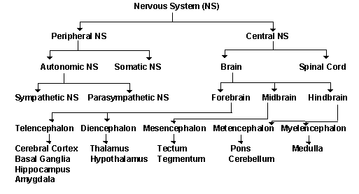
·
The
Central Nervous System (CNS), which
consists of the brain and the spinal cord. It receives, processes, stores and
transfers information.
·
The
Peripheral Nervous System (PNS)
includes the components of the nervous systems that lie outside the CNS
The PNS has two functional subdivisions (Johnson,
2012):
1.
The
sensory division of the PNS carries
information to the brain and spinal cord
2.
The
motor division carries information
from the CNS to other parts of the body.
The motor division of the PNS is subdivided along
functional lines:
·
The
somatic division controls skeletal muscles
·
Autonomic
division
controls smooth muscles, cardiac muscles and glands
The Autonomic division has two subdivisions:
1.
Sympathetic
division
2.
Parasympathetic
division
These two
divisions work against each other to accomplish the automatic, subconscious
maintenance of homeostasis.
The nervous system is the body’s main
control system that receives input from a variety of sources. It controls our
body’s physical movements, maintains homeostasis of many internal variables and
initiates our higher thought processes and emotions.
11.2 Neurons are
the communication cells of the nervous system
Neurons are specialized cells for communication
(Oellers, 2012). They generate and conduct electrical impulses called action
potentials from one part of the body to another. The longest neurons extend all
the way from your toes to your spinal cord.
There are three types of neurons (Oellers, 2012):

https://encrypted-tbn3.google.com/images?q=tbn:ANd9GcRJJYtIICpQHDUQKfvKTU-GcQqvefQbxxLonPW9CduS2NPORr8nvA, Accessed May 4, 2012.
1.
Sensory neurons found in the
PNS that receive stimuli and transmit information to the CNS.
2.
Interneurons transmit
information between components of the CNS.
3.
Motor neurons found in the
PNS transmit information away from the CNS.

http://www.emc.maricopa.edu/faculty/farabee/biobk/neurons_1.gif, Accessed May
4, 2012.
All neurons
consist of a cell body (main body), one or more dendrites (extensions of the
cell body) and an axon. The cell body
contains the nucleus with its DNA, mitochondria and other cell organelles. Dendrites receive information from
receptors or incoming impulses from other neurons. An axon is a long, slender tube of cell membrane containing a small
amount of cytoplasm. Axons conduct electrical impulses. The axon branches into
slender extensions called axon terminals.
Each axon terminal ends in a small, rounded tip called an axon bulb. The flow
of information begins at a receptor near a dendrite ending of a sensory neuron
and ends at the axon bulbs of a motor neuron. Sensory neurons transmit impulses
to the CNS. Interneurons transmit impulses between components of the CNS. Motor
neurons transmit impulses away from the CNS to muscles and glands.
11.3 Neurons
initiate action potentials
Neurons function
is to transmit information from one part of the body to another in the form of
electrical pulses. How neurons communicate with one another is by chemically.
Information passes from one neuron to another by the release of chemical
substances called neurotransmitters.
Neurons initiate action potentials,
which are electrical impulses, based on the movement of ions in and out of the
cell membrane of the neuron (Oellers, 2012). Neurons generate and transmit
action potentials.
11.3a
Sodium-potassium pump maintains resting potential
The sodium-potassium pump is essential for
the development and maintenance of an electrical charge across the cell
membrane (Johnson, 2012). This is because the sodium-potassium pump has a difference
in charges by transporting positive sodium ions out of the cell from every two
potassium ions transported in. The sodium-potassium pump is removing both
osmotic particles and positive charges from the cell at the same time resulting
in the presence of negatively charged protein molecules that are trapped within
the cell. This result in an electrical charge across the cell membrane called
the membrane potential.

http://www.au.dk/fileadmin/www.au.dk/forskning/nobelprisen_i_kemi_1997/natrium-kalium-pumpen/cellenuk.jpg, Accessed May
4, 2012.
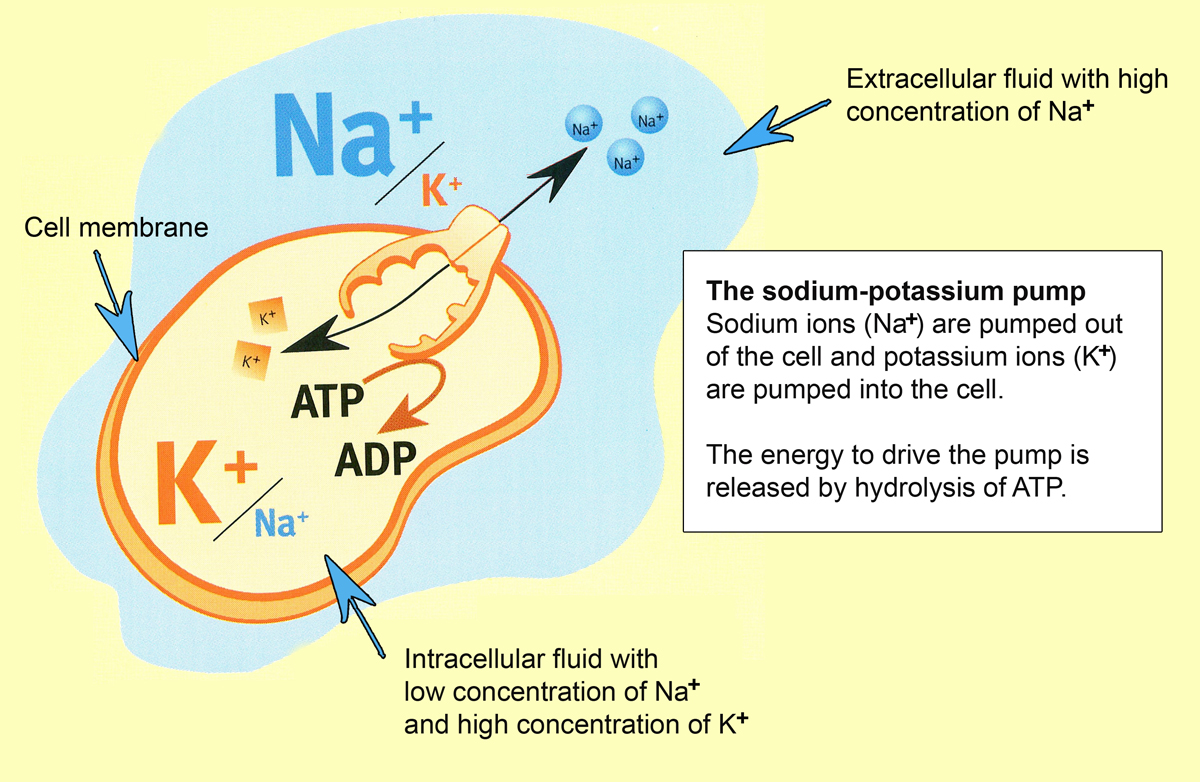
The normal
membrane potential at rest is its resting
(membrane) potential. This means that the inside of the cell is a slightly
different charge than the outside (Oellers, 2012). Resting potential measures
the difference in voltage across the cell membrane in a resting cell.

http://www.ncbi.nlm.nih.gov/books/NBK9847/bin/ch12f20.jpg, Accessed May
4, 2012
Functions of the Sodium-Potassium Pump (Oellers,
2012):
·
Maintains
cell volume
·
Establishes
and maintains resting potential by active transport of sodium out of the cell
and potassium in.
The
concentration of sodium is much higher in the interstitial fluid than it is in
the cytoplasm. Because sodium is always leaking into the cell and potassium is
leaking out by passive diffusion balances the rate of leakage. Not only is potassium
important for action potentials, but researchers have found that potassium is
linked to memory and learning. ScienceDaily 2012, state in their article that
scientists discovered that mice missing the channel that delivers potassium
also showed diminished learning abilities. Scientists are hoping that by
targeting the chemical pathways that alter potassium channels they may be able
to apply the findings to humans and reverse some of the cognitive deficits in
people with epilepsy and other neurological disorders (ScienceDaily 2012).
11.3b Graded
potentials alter the resting potential
Every time an
impulse arrives from a neuron the resting potential changes. Depending on the
type of signal and its strength the change might depolarize the membrane (move the voltage closer to zero) or hyperpolarize it (make it even more
negative). These changes in the resting potential are called graded potentials because they can vary
in size. Graded potentials fade away at increasing distances from a single
region on the membrane (Johnson, 2012). Summation
occurs when many incoming signals from other neurons produce a bigger change in
the resting potential.
11.3c An action
potential is a sudden reversal of membrane voltage
An action
potential is initiated when graded potentials reach a certain threshold
(triggering point) (Oellers, 2012). After the threshold is reached there is a
sudden temporary reversal of the voltage difference across the cell membrane.
Once this action potential is initiated, it sweeps rapidly down the axon at constant
amplitude and rate of speed until it reaches the axon terminals. An action
potential impulse is the only form in which information is transmitted long
distance by the nervous system. Action potentials occur because the axon
membrane contains voltage-sensitive ion channels that open and close sequentially
once threshold is reached.

Picture of an
Action Potential
http://faculty.washington.edu/chudler/ap3.gif, Accessed May
4, 2012.
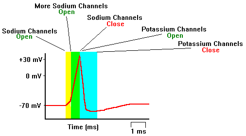
An action potential happens in a sequence of three
events (Johnson, 2012):
1.
Depolarization happens when
the threshold is exceeded, sodium channels open briefly and the membrane
permeability to sodium increases. Sodium diffuses rapidly into the axon, depolarizing
the membrane (the membrane potential turns from negative to positive).
2.
Repolarization is when sodium
channels close. Potassium channels open and the membrane permeability to
potassium increases. Potassium diffuses outward, repolarizing the membrane (the
interior of the axon becomes negative again).
3.
Reestablishment of the resting
potential happens after a brief hyper polarization caused by a delay in the
full closure of potassium channels, the membrane potential returns to its
normal resting value.
While an action
potential is under way, an axon cannot generate another action potential this
is called the absolute refractory period
this ensures the action potential always travel in one direction only.
The absolute
refractory period is followed by a brief relative refractory period during
which it is harder than usual to generate the next action potential.
11.3d Action
potentials are all-or-none and self-propagating
action potential does not occur unless an
individual neuron threshold sets the extent of the stimulus needed (Oellers,
2012). If it achieves the threshold, it fires and an action potential occurs.
The form and voltage of the action potential are always the same no matter how high
the graded stimulus is. An action potential is “all-or-none” either it occurs or it does not.
An action
potential is self-propagating it multiplies
itself into the next region of the axon. It moves like a wave down the axon,
with constant speed and amplitude (Oellers, 2012).
Neurons transmit
information about stimulus intensity by the number of action potentials that
are being generated and transmitted per unit time. Stronger stimuli generate
more action potentials and unit time. The speed of an action potential is
always constant for a given neuron. Action potential speed is greater in
larger-diameter axons and in axons covered by an insulating sheath.
11.4 Neuroglial
cells support and protect neurons

http://www.mananatomy.com/basic-anatomy/glial-cells-neuroglia,
Accessed May 4, 2012.
80% of cells in the nervous system are neuroglial cells (Oellers, 2012). These
cells provide physical support and protection to neurons and help maintain
healthy concentrations of important chemicals in the fluid surrounding them.
These cells do not generate or transmit impulses.

http://images.sciencedaily.com/2005/09/050902073203.jpg, Accessed May
4, 2012.
Schwann cells produce a fatty insulating material
called myelin. This material
encloses and protects many neuron axons in the PNS peripheral nervous system. A
myelin sheath is created when an
individual Schwann cell during development wraps itself around a short segment
of an axon many times, creating a shiny white protective layer around the axon.
Nodes of Ranvier are between adjacent
Schwann cells and are short non-insulated gaps where the surface of the axon is
still exposed. Myelinated neurons
are neurons that have axons wrapped in a sheath of myelin.
The myelin sheath serves three important functions
(Johnson, 2012):
1.
It
saves the neurons energy. The insulating layer of the myelin prevents some of
the inward leak of sodium and outward leak of potassium. These leaks normally
have to be replaced by active transport processes requiring energy.
2.
It
seeds up the transmission of impulses. The myelin sheath prevents all leakage
of charged ions across the axon membrane except where the axon is bare (nodes
of Ranvier). The local depolarizing current spreads much farther within the
axon all the way to the next node of Ranvier, making the rate of travel even
faster than the rate of travel of a continuously propagated action potential
along an unmyelinated axon.
3.
It
helps damaged or severed axons of the peripheral nervous system regenerate. If
a neuron axon is severed, it can grow back because it is still attached to the
cell body by the sheath.
Oligodendrocyte is another type of neuroglial
cell that produces protective sheaths of myelin in the CNS. Oligodendrocytes
degenerate after injury unlike the Schwann cells. This is why spinal cord
injuries and CNS disorders such as MS multiple sclerosis result in a permanent
change or loss of function. People with MS can suffer a variety of symptoms
depending on which areas of the CNS are damaged. Common symptoms include muscle
weakness, visual impairment and urinary incontinence. MS is progressive disease
(Johnson, 2012).
11.5 Information
is transferred from a neuron to its target
In order for neurons
to communicate with each other, a chemical substance called a neurotransmitter must be released that
crosses between two cells called a synapse.
The process of transmission from a neuron to its target is called synaptic transmission. Targets can be
another neuron, muscle cell or gland (Oellers, 2012).
11.5a
Neurotransmitter is released
http://academic.uprm.edu/~ephoebus/07b80e00.gif, Accessed May 4, 2012.
At a synapse,
the presynaptic membrane is the cell
membrane of the neuron that is sending the information. The postsynaptic membrane is the membrane of the cell that is about to
receive the information. The synaptic
cleft is the small fluid-filled gap that separates the two.
The events that occur during a synaptic transmission
occur as follows (Oellers, 2012):
·
An
action potential arrives causing calcium to diffuse into the axon bulb
·
Calcium
causes the release of neurotransmitters from vesicles.
·
Neurotransmitter
diffuses across synaptic cleft, binds to receptors on target (postsynaptic)
membrane, and opens gated channels.
·
Graded potential results from sodium movement
through opened channels
11.5b
Neurotransmitter exert excitatory or inhibitory effects
The response of
the postsynaptic cell to neurotransmitter depends on several factors, including
the type of neurotransmitter, type of receptors and type of gated ion channels
(Oellers, 2012).
Neurotransmitters are classified as (Johnson, 2012):
·
Excitatory
neurotransmitters
that depolarize the postsynaptic cell causing threshold to be exceeded or
approached and the generation of new impulses in the postsynaptic neuron.
·
Inhibitory neurotransmitters cause the
postsynaptic cell to hyperpolarize and make it harder for threshold to be reached.
These neurotransmitters prevent the generation of action potentials in the
postsynaptic neuron.
·
Some
neurotransmitters can be excitatory or inhibitory depending on the type of
receptor to which they bind on the postsynaptic membrane.
Prompt removal
of neurotransmitter causes neural signals to be terminated rapidly. Only then
can the next message be received.
The neurotransmitter may be removed from the
synaptic cleft in three ways (Johnson, 2012):
1.
It
may be taken back up again by the presynaptic neuron and repackaged into
membrane-bound vesicles, to be used again
2.
It
may be destroyed by enzymes in the synaptic cleft
3.
It
may diffuse away from the synaptic cleft into the general circulation, where it
will ultimately be destroyed
11.5c
Postsynaptic neurons integrate and process information
Postsynaptic
neurons may integrate incoming signals from many different presynaptic neurons.
The conversion of the signal from electrical (action potential) to chemical
(neurotransmitter) allows the postsynaptic cell to do a lot of integration and
information processing. One way for threshold to be reached in a postsynaptic
neuron is for the presynaptic neuron to increase frequency of stimulation,
sending lots of action potential in a short time. Response in postsynaptic cell
depends on how many neurons are forming synapses with it and wheter the neurons
forming synapses are excitatory or inhibitory (Oellers, 2012). One neuron
receives input from many neurons called convergence.
Divergence occurs when one neuron sends action potentials to multiply other
neurons.
REFERENCES
Johnson,
M. D. (2012, 2010, 2008). Human Biology: concepts and current issues, sixth edition.
Pearson Education, inc.; Benjamin Cummings.
Oellers, J. (n.d). Online
Presentation: Ch11. The Nervous System: Integration and Control. Retrieved May
5, 2012, from http://lblackboard.yc.edu/webapps/portal/frameset.jsp?tab_tab_group_id=_2_1&url=%2Fwebapps%2Fblackboard%2Fexecute%2Flauncher%3Dcourse%26id%3D_43466_1
Science Daily (April 26, 2012).
Clues to Reverse Cognitive Deficits in People with Neurological Disorders.
Retrieved May 5, 2012, from
Ch. 12 Sensory
Mechanisms
Table of Contents
12.1 Receptors receive and convert stimuli
a. Receptors are classified
according to stimulus
b. The CNS interprets nerve impulses
based on origin and frequency
c. Some receptors adapt to
continuing stimuli
d. Somatic sensations and special
senses provide sensory information
12.2 Somatic sensations arise from receptors
throughout the body
a. Mechanoreceptors detect touch,
pressure and vibration
b. Mechanoreceptors indicate limb position,
muscle length and tension
c. Thermoreceptors detect
temperature
d. Pain receptors signal discomfort
12.6 Vision: Detecting and interpreting visual
stimuli
a. Structure of the eye
b. Regulating the amount of light
and focusing the image
c. Eyeball shape affects focus
d. Light is converted into action
potentials
e. Rods and cones respond to light
f. Rods provide vision in dim light
g. Cones provide color vision and
accurate images
h. Visual receptors adapt
12.7 Disorders of sensory mechanisms
a. Disorders of the ears
1. Deafness: loss of
hearing
2. Otitis media:
Inflammation of the middle ear
3. Meniere’s syndrome:
Inner ear condition impairs hearing and balance
b. Disorders of the eyes
1. Retinal detachment:
Retina separates from choroid
2. Cataracts: The lens
become opaque
3. Glaucoma: Pressure
inside the eye rises
4. Age-related macular
degeneration
5. Color blindness:
Inability to distinguish the full range of colors
12.1 Receptors
receive and convert stimuli
Stimulus is sensory input that causes a change
within or outside the body (Johnson 2012). The stimulus is often a form of
physical energy such as heat, pressure, or sound waves, but it can also be a
chemical. Receptors are located throughout the body and provide us with
information about body position, touch, temperature, vibration, pressure and
pain.
Some receptors
convert the stimulus into a graded potential that, if powerful enough, initiate
an impulse within the sensory neuron. Other receptors are parts of cells that
produce graded potentials and release a neurotransmitter, stimulating nearby
sensory neuron.
We feel
sensations when the CNS receives impulses and we perceive these impulses as
what we understand the sensations are. For example, when we hear thunder we
know that a storm is on its way.
12.1a Receptors
are classified according to stimulus
Receptors are classified according to type of
stimulus energy they convert (Johnson 2012):
·
Mechanoreceptors
respond
to forms of mechanical energy, such as waves or sound, changes in fluid
pressure, physical touch or pressure, stretching or forces generated by gravity
and acceleration.
·
Thermoreceptors respond to heat
or cold.
·
Pain receptors respond to
tissue damage or excessive pressure or temperature.
·
Chemoreceptor’s respond to the
presence of chemicals in the nearby area
·
Photoreceptors respond to
light.
Many receptors
contribute to sensations and some to giving us a sense of where our limbs are
located. Stretch receptors monitor
and regulate blood pressure and fluid volumes, and chemoreceptor’s regulate the chemical composition of our internal
environment. These receptors are “silent” we are not consciously aware of their
actions and they function in negative feedback loops that maintain homeostasis
inside the body.
12.1b The CNS
interprets nerve impulses based on origin and frequency
Cells in the CNS
interpret incoming impulses as images because nerve impulses are transmitted
from receptors to specific brain areas. Impulses generated by visual stimuli
travel in sensory neurons whose axons go directly to brain regions associated
with vision. All incoming impulses traveling in these neurons are interpreted
as light. Cells in the CNS are also able to distinguish a loud sound from a
soft one. Stronger stimuli activate more receptors and trigger a greater
frequency of impulses in sensor neurons.
12.1c Some
receptors adapt to continuing stimuli
The CNS can
ignore one sensation to concentrate on others. For example, if you are
interested in something else, you do not focus on how your clothes feel on your
body. Some sensory inputs are ignored after a while, because of receptor adaptation. This is where the
sensory neuron stops sending impulses even though the original stimulus is
still present. For example touch a strand of your hair, you will feel an initial
sensation and then that sensation disappears.
There are
receptors in the skin for light touch and pressure. This is for a survival
advantage because they keep the CNS informed of changes in the stimuli, without
bombarding it with relatively unimportant stimuli (Johnson 2012). Olfactory are smell receptors that
inform us of different smells. Other receptors include pain, joint and muscle
receptors that are used to monitor the position of our limbs, and all of the
silent receptors involved in homeostatic feedback control loops. Some receptors
adapt rapidly while others adapt slowly or not at all.
12.1d Somatic
sensations and special senses provide sensory information
Senses (sensations) provided by receptors are
categorized as (Johnson 2012):
·
Somatic sensations originate from receptors
present at more than one location in the body. These include temperature,
touch, vibration, pressure, pain and awareness of body movements and position.

https://encrypted-tbn1.google.com/images?q=tbn:ANd9GcQCV2808B3ze1iKz5xceMDFudhQG1BOcUikqX5OMJ4NNUxOLHVxgQ,
Accessed May 7, 2012.
·
Special senses originate from
receptors that are restricted to particular areas of the body, such as the ears
and eyes. They deliver highly specialized information about the external world.
12.2 Somatic
sensations arise from receptors throughout the body
Somatic sensations are essential to help us
coordinate muscle movements, avoid danger, and maintain body temperature. These
sensations are also the response we have when we are touched by a loved one.
Receptors that
detect the somatic sensations are located throughout the body in skin, joints,
skeletal muscles, tendons and internal organs (Johnson 2012). Sensory neurons
linked to these receptors send their impulses to the brain, specifically to the
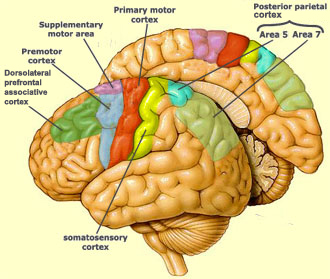
primary somatosensory area of the parietal love of the cerebral cortex. Parts
of the body with the greatest sensory sensitivity such as the mouth and fingers
involve more neurons in the somatosensory area, where less sensitive body parts
involve fewer neurons.

http://thebrain.mcgill.ca/flash/a/a_06/a_06_cr/a_06_cr_mou/a_06_cr_mou_1a.jpg,
Accessed May 7, 2012.
The somatosensory area processes the
information and sends it to the nearby primary motor area in the frontal lobe.
If necessary, impulses are then generated in motor (output) neurons of the
peripheral nervous system to cause body movement.
12.2a
Mechanoreceptors detect touch, pressure, and vibration
Mechanoreceptors are receptors for sensing touch,
pressure and vibration. These receptors may take several different forms but
all mechanoreceptors are the modified dendrite endings of sensory neurons. Any
force that deforms the plasma membrane of the dendrite ending produces a
typical graded potential. If the graded potential is large enough to exceed
threshold, the sensory neuron initiates an impulse.
The various
receptors for touch, pressure and vibration differ in location.
Vibration-sensitive receptors adapt quickly and take a rapidly changing
physical deformation to keep them stimulated. These types of receptors are
useful for providing information about insects flying around us.
Several types of
receptors for detecting somatic sensations as follows (Johnson 2012):

http://bdml.stanford.edu/twiki/pub/Haptics/ProjectOverview/mechanoreceptors.jpg,
Accessed May 7, 2012.
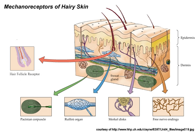
·
Unencapsulated
dendritic endings
are naked dendrite endings of sensory neurons around hairs and near the skin
surface to signal pain, light pressure, and changes in temperature.
·
Merkel disks are modified
unencapsulated dendritic endings that detect light touch and pressure.
·
Meissner’s
corpuscles
are encapsulated touch receptors located close to the skin surface that detect
the beginning and the end of light pressure and touch.
·
Ruffini endings are
encapsulated receptors that respond continually to ongoing pressure.
·
Pacinian
corpuscles
consists of several kinds of dendrite endings are encapsulated (enclosed) in
epithelial or connective tissue. The Pacinian corpuscles are enclosed receptors
located in the dermis that respond to either deep pressure or high-frequency
vibration. This receptor permits the mechanoreceptor to adapt rapidly, even to
a continuously applied stimulus.
12.2b
Mechanoreceptors indicate limb position, muscle length and tension

http://faculty.pasadena.edu/dkwon/PNS%20and%20propioception/peripheral%20nervous%20system%20and%20propioception_files/images/image29.png,
Accessed May 7, 2012.
A variety of
Mechanoreceptors in our joints (for joint position), skeletal muscle (for
muscle length), and in tendons (for tension) make it possible for us to close
our eyes and know where our limbs are or make it easy for us to identify which
muscle groups are contracting. Muscle
spindles are mechanoreceptors that monitor muscle length. For most joints,
muscle length determines joint position because of the way the muscle is
attached to the bones. Muscle spindles are small bundles of modified skeletal
muscle cells located within a skeletal muscle. Sensory nerves whose dendritic endings are mechanoreceptors are
attached to the muscle spindles. When the whole muscle is stretched, so do the
receptors that are attached. Mechanical
distortion of the mechanoreceptors produces local graded potentials in the
dendrite endings and an action potential. Example of this action is a reflex. Tendons, which connect muscle to bone,
have mechanoreceptors that respond to tension, but they cannot distinguish
between tension produced by passive stretch and tension produced by active
muscle contraction. Tendon receptors
help protect you from injury by extremely high tensions. An example of this is
falling out of tree and when you hit the ground, you automatically fall. This
effect is a reflex induced collapse
that protects your muscles from tearing and your bones from breaking.
12.2c Thermoreceptors
detect temperature

http://www.exploringnature.org/graphics/anatomy/sensory%20organs.jpg,
Accessed May 7, 2012.
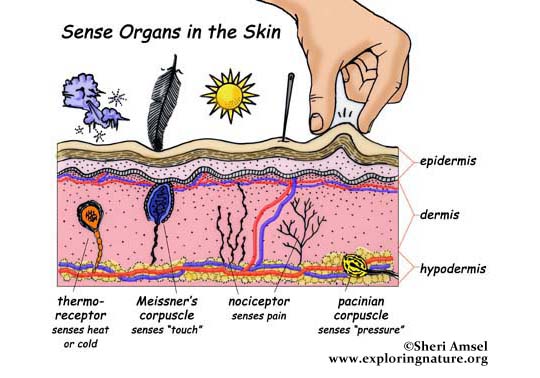
Thermoreceptors are near the skin’s surface and
detect heat and cold, which provide information about the external environment
(Johnson 2012). They adapt quickly, allowing us to monitor changes in temperature
accurately and adjust sensory input so it becomes more bearable. An example of
this is swimming in a lake. At first, the water is cold but soon your body
adjusts to the temperature and you no longer feel cold.
Other
thermoreceptors are located in the abs and chest organs throughout the body
that monitor internal temperatures (core temperature). They help in maintain
homeostasis and they do not adapt quickly.
12.2d Pain
receptors signal discomfort
Pain receptors are unencapsulated endings that
respond to injury from excessive physical pressure, heat, light or chemicals.
Pain can be depending on its characteristics
(Johnson 2012):
·
Fast
·
Slow
Fast pain can be either acute or sharp and occurs
quickly after the stimulus. Receptors for fast pain respond to physical
pressure or heat and usually are located near the surface of the body. They
inform us of stimuli to be avoided, for example, a hot burner on a stove that
when we touch we automatically draw our hand away; this is in response to fast
pain that is rapid and strong.
Slow pain arises from muscles or internal organs.
This type of pain is due to activation of chemically sensitive pain receptors
by chemicals released from damaged tissue. Slow pain from internal organs is
often perceived as originating from an area of the body completely removed from
the actual source. This is called referred
pain. This happens because action potentials from internal pain receptors
are transmitted to the brain by the same spinal neurons that transmit action
potentials from pain receptors in the skin and skeletal muscles. The brain has
no way of knowing the exact source of the pain so it assigns the pain to
another location. An example of this is someone who is having a heart attack;
you will initially feel the pain in the left shoulder and down the left arm.
Pain receptors do not adapt for survival purposes.
12.6 Vision:
Detecting and interpreting visual stimuli
Light is a form
of electromagnetic radiation (Johnson 2012). Our eyes allow us to receive and
process light. They enable us to detect light from objects both nearby and
distant and from sources either dim or bright. Photoreceptors are cells in our eyes that collect what we see and
focus it.
12.6a Structure
of the eye

http://www.anatomy.tv/StudyGuides/Images/eyeball2.jpg,
Accessed May 7, 2012.
A tough outer
coat known as the sclera (white of
the eye) covers the outer surface except in the very front, where it is
continuous with the clear cornea.
Light enters
through the transparent cornea and
passes through aqueous humor (that
nourishes and cushions the cornea and lens). Light then either strikes the iris (colored disk-shaped muscle that
determines how much light enters the eye or passes through the pupil (adjustable opening in the center
of the iris). The light then strikes the lens
(a transparent, flexible structure attached by connective tissue fibers to a
ring of circularly arranged smooth muscle called the ciliary muscle). Light
then passes through the main chamber of the eye filled with vitreous humor.
Light encounters the layers at the back and sides of the eye (Johnson 2012).
This is the retinal (primarily
photoreceptor cells, neurons and a few blood vessels) and the the back of the
eye where the choroid (comprises of pigmented
cells and blood vessels). The pigmented
cells absorb light not sensed by photoreceptors so that the image is not
distorted by reflected light and the blood vessels nourish the retina (Johnson
2012). At the back of the eye is the optic
nerve, which carries information to the thalamus to be forwarded to the visual cortex for interpretation. Finally,
skeletal muscles surround the eye
and control its movements so we can choose where to look. The macula is the central region of the
retina where photoreceptor density is the highest. When we want to focus in on
an object, we look directly at it. At the very center of the macula is a small
pit called the fovea centralis that
is lined with photoreceptors (Johnson 2012). The optic disk is where the axons of the optic nerve and blood vessels
exit the eye, so there are no photoreceptors. The optic disk leaves us with a
“blind spot” in each eye.
12.6b Regulating
the amount of light and focusing the image
The iris adjusts the amount of light
entering the eye with two sets of smooth muscles. Bright lights would overwhelm
our photoreceptors and blind us if the muscles in our eyes did not cause our
pupil to contract. In dim light, contraction of smooth muscles arranged
radially around the pupil causes the pupil to dilate (Johnson 2012).
Nerves control
each set of muscles. To test this, physicians will shine a flashlight into your
eyes. Your pupils should be small in response to the light and when the light
is removed, the pupils should get bigger.
The cornea and the lens focus light entering the eye. The cornea is curved and not adjustable.
It is responsible for bending most of the incoming light. However, changing the
curvature of the lens regulates the intensity of the incoming light, changes
our focus between near, and far objects.
To focus on an
object that is near the ciliary muscle will contract and the inner radius of
the muscle shrinks reducing the tension of the fibers attached to the lens
(Johnson 2012). This allows the lens to focus on an abject that is near. When
the ciliary muscle relaxes, the ring of muscle increases the tension on the
lens, stretching and flattening it and bringing objects that are more distant
into focus. Accommodation refers to
the adjustment of lens curvature so we can focus on near or far objects.
The light rays
from each point of an object are bent and focused so that the image created on
the retinal is upside down (inverted). The brain interprets the image as right
side up.
Around forty
years old our lens will stiffen and cannot resume a bulging shape even when the
ciliary muscles are contracted (Johnson, 2012). This results in presbyopia, the inability to focus on nearby
objects.
12.6c Eyeball
shape affects focus

http://www.eyecarecontacts.com/optical_lenses.GIF,
Accessed May 7, 2012.
Differences in
the shape of the eyeball affect our ability to focus properly. Myopia is a common inherited condition
in which the eyeball is slightly longer than normal (Johnson, 2012). Even when
the lens is flattened, distant objects focus in front of the retina. People
with this disorder can see nearby objects, but objects far away are blurry.
This is called nearsightedness. Concave lenses bend incoming light so
it focuses on the retina. This can correct nearsightedness.
Hyperopia (farsightedness) is a disorder
where people can see objects far away, but objects close by are blurry (Johnson
2012). This occurs when the eyeball is too short and nearby objects focus
behind the retina. Convex lenses can correct this. Astigmatism is blurred vision caused by irregularities of the shape
of the cornea or lens. This results in light being scattered and may not focus
evenly on the retina. Specialty ground lenses that exactly compensate for the
irregularities of the cornea and lens can fix this.
12.6d Light is
converted into action potentials
The eyes convert
stimulus (light) into impulses. This process occurs in the retina. The retinal allows us to see in color,
adapt light intensities, and perceive images of the world around us.
The retina consists of four layers (Johnson 2012):

http://www.saburchill.com/IBbiology/chapters03/images/18050312.jpg,
Accessed May 7, 2012.
1.
The
outermost layer consists of pigmented cells that along with the choroid absorb
light not captured by the photoreceptor cells.
2.
Next
layer of photoreceptor cells are called rods
and cones because of their
shape.
3.
The
rods and cones synapse with the third layer of neurons called bipolar cells. These cells process and
integrate information and then pass it on to the fourth layer.
4.
The
innermost layer consists of ganglion
cells. These cells are neurons with long axons that become the optic nerve
going to the brain.
12.6e Rods and
cones respond to light
The rods and
cones shape, consist of a series of flattened disks arranged to form a rod or
cone shaped structure at one end. The flattened disks contain molecules of a light-sensitive
protein called photopigment (Johnson
2012). This protein undergoes a change of shape when exposed to light. This
change of shape causes the photoreceptors (rod or cone) to close some of its
sodium channels and reduce the amount of neurotransmitter it normally releases.
This activates the ganglion cells.
12.6f Rods
provide vision in dim light
Rods all have
the same photopigment called rhodopsin. Rhodopsin
is much more sensitive to light than the photopigments in cones. In dim light,
our vision is dependent on rods because of this. Rods do not give us color vision,
which is why objects appear less colorful in dim light (Johnson, 2012). Rods
and cones are not distributed evenly on the retina. Regions of the retina farthest
away from the fovea have the highest ration of rods to cones.
12.6g Cones
provide color vision and accurate images
We are able to
see colors because we have three different kinds of cones: red, green and blue
(Johnson 2012). Each contains a photopigment that absorbs the light of red,
green or blue light. Our ability to distinguish a variety of colors is due to
the way the brain interprets the ratios of impulses coming from the ganglion
cells connected to the three types of cones. When all three types are activated
by different wavelengths, we see white light. When we see black, it is because
there is an absence of light. Cones require stronger light to be activated
because the cone photopigments are much less sensitive to light than the
rhodopsin in rods. This is why our ability to distinguish between colors
declines in dim light. In dim light, you see with your rods, which can detect
only black and white.
12.6h Visual
receptors adapt
Vision adapts to
changing light conditions over several seconds or minutes. Adaptation takes
longer when going from bright to dim light than it does in the opposite
direction. Adaptation depends on rapid adjustment of the pupil by the iris and
on adaptation by the rods.
Rhodopsin
absorbs light and uses up the photopigment temporarily. Light energy breaks the
rhodopsin into two molecules, which can be resynthesized into rhodopsin again
over a period of minutes. Being in bright light for long periods causes most of
your rhodopsin to be broken down. Then when you enter a dim room your cones are
no longer working and there is not enough intact rhodopsin for you to see very
well in the dim room. With time, your rhodopsin is resynthesized and your
vision becomes clear. When you go outside on a bright day from a dim room the
light seems very bright because you have the maximum amount of photopigment in
both rods and cones.
12.7 Disorders
of sensory mechanisms
There are many
disorders of your senses. We will focus on disorders concerning the ears and
eyes.
12.7a Disorders
of the ears
Having problems
hearing can range from having a loss of hearing-to-hearing impairment.
Scientists have discovered that that hearing is also linked to touch. People
with good hearing also have a keen sense of touch; people with impaired hearing
generally have an impaired sense of touch (ScienceDaily 2012). ScienceDaily
2012 reports in their article that scientists during a five year experiment
have discovered that women hear better than men and feel more.
12.7a, 1
Deafness: loss of hearing

http://media-3.web.britannica.com/eb-media/04/14304-004-6C1B7EB1.gif,
Accessed May 7, 2012.
Deafness is caused by damage to hair cells
(Johnson, 2012). This is called nerve deafness because sounds can be converted
into impulses in sensory nerves. Nerve deafness results from frequent exposure
to loud sounds. Damage to the bones of the middle ear is called conduction deafness. Sound waves are
not transferred to the inner rear at all. This is caused by arthritis of the
middle ear bones. A hearing aid can correct some types of hearing loss. If hair
cells are missing, however the amplification of a hearing aid will not be
effective. A tiny implanted microprocessor can help with this. This device
converts sound waves into electrical signals.
12.7a, 2 Otitis
media: Inflammation of the middle ear
An earache is
caused by otitis media, an
inflammation of the middle ear that results from an upper respiratory tract
infection (Johnson 2012). This tract infection extends up the auditory tube and
can become blocked and trapped with fluid. Antibiotics can treat this.
12.7a, 3
Meniere’s syndrome: Inner ear condition impairs hearing and balance
Menieres syndrome is a chronic condition of the
inner ear. The cause is unknown but can be excess fluid in the cochlea and
semicircular canals. Symptoms include dizziness and nausea by progressive
hearing loss. Balance can be affected finding it difficult to stand upright.
Mild cases can be treated with motion sickness medications. Severe cases may
benefit from surgery to drain excel fluid from the inner ear.
12.7b Disorders
of the eyes
Disorders of the
eyes can include blindness, cataracts, glaucoma, visual impairment and color
blindness.
12.7b, 1 Retinal
detachment: Retina separates from choroid
A blow to the
head causes retinal detachment. What
happens is that the retina is torn and the vitreous humor leaks through the tear
and peels the retina away from the choroid (Johnson 2012). The detached region
of the retina loses most of its blood supply and its ability to focus on an
image properly. Symptoms include flashes of light, blurred vision or loss of
peripheral vision. Prompt surgery can usually repair the damage.
12.7b, 2
Cataracts: The lens becomes opaque

https://encrypted-tbn0.google.com/images?q=tbn:ANd9GcQIcraqcAIf_3xA1-wxVKAVWUwOhxj5bwhcvjaM08c34ZoY5glR5g,
Accessed May 7, 2012.
Cataracts are a
decrease in the normal transparency of a lens. Normally the lens contains a
kind of protein that is folded in such a way that it is transparent. If the delivery
of nutrients is inadequate these proteins may denature or clump making the lens
opaque (not allowing the light to pass through the retina) (Johnson 2012). Some
cataracts are present at birth (congenital), but most are age-related.
Treatment includes removing the lens surgically and replacing it with an artificial
lens.
12.7b, 3
Glaucoma: Pressure inside the eye rises
Glaucoma is a condition in which the drainage
vessel that drains the aqueous humor (brings nutrients and oxygen to the lens
and cornea and carries away wastes) become blocked (Johnson 2012). The excess
fluid increases pressure inside the eye and compresses blood vessels supplying
the retina. Cells of the retina or optic nerve may die, impairing vision and
often will start as a loss of peripheral vision. Glaucoma generally develops
slowly and painlessly over several years without any symptoms. Most people do
not realize they have Glaucoma until their vision starts to fade. Although not
common, but glaucoma can develop acutely (within hours) with symptoms of
blurred vision and red, swollen and sometimes painful eyes. Acute glaucoma requires
immediate medical care. Glaucoma can be detected with a test that measures the
pressure in the eye. Glaucoma if detected early can be control with drugs or
surgery before permanent damage occurs. However, any vision that has been lost
cannot be restored.
12.7b, 4
Age-related macular degeneration

http://maculardegenerationdisease.com/wp-content/uploads/2010/10/Different-types-of-Macular-Degeneration.jpg,
Accessed May 7, 2012.
Age-related macular degeneration is a disease of
visual impairment caused by detachment of the retinal and degeneration of
photoreceptor cells in the macular region of the retina (Johnson 2012). The
most common cause of AMD is accumulation of cellular debris between the choroid
and the retina. AMD can also be caused by an abnormal growth of blood vessels
in this region. AMD results in a loss of visual acuity in the center of the
visual field making it difficult to recognize faces or to read. AMD is the
leading cause of loss of vision in persons over 60. There is no effective cure.
12.7b, 5 Color
blindness: Inability to distinguish the full range of colors
Deficient
numbers of particular types of cones causes most forms of color blindness. One
of the three cones can also be missing. People with red-green color blindness
are deficient in either red cones or green cones. They have trouble
distinguishing between red and green or they can perceive them as the same
color. The inability to perceive any color at all is caused when two of the
three cones are missing. Color blindness if often inherited. Red-green color
blindness is an X-linked recessive trait that is more common in men than women
are. Color blindness can be tested with a series of colored test plates.
REFERENCES
Johnson,
M. D. (2012, 2010, 2008). Human Biology: concepts and current issues, sixth
edition. Pearson Education, inc.; Benjamin Cummings.
Science Daily (May 1, 2012). Hearing
and Touch Have Common Genetic Basis: Gene Mutation Leads to Impairment of Two
Senses. Retrieved May 6, 2012, from
Ch. 24 Human
Impacts, Biodiversity, and Environmental Issues
Table of Contents
24.1 Pollutants impair air quality
a. Excessive greenhouse gases lead
to global warming
b. CFCs deplete the ozone layer
c. Pollutants produce acid precipitation
d. Smog blankets industrial areas
24.2 Pollution jeopardizes scarce water supplies
a. Water is scarce and unequally
distributed
b. Urbanization increases storm
water runoff
c. Human activities pollute
freshwater
d. Groundwater pollution may impair
human health
e. Oil pollution damages oceans and
shorelines
24.3 Pollution and overuse damage the land
24.1 Pollutants impair
air quality
Air consists of
79% nitrogen, 21% oxygen with trace amounts of carbon dioxide (Johnson, 2012).
Pollution is also found in the air we breathe. Thousands of chemicals and
particles have adverse effects on every living organism on Earth.
The concerns of
air pollution fall into four areas (Johnson, 2012):
1.
Global
warming
2.
Destruction
of the ozone layer
3.
Acid
precipitation
4.
Smog
production
Air pollution is
hard to get rid of because there are so many possibilities to where the
pollution came from.
24.1a Excessive
greenhouse gases lead to global warming
Greenhouse gases are gases that are caught in
Earth’s atmosphere and increase the temperatures on Earth, like a green house.
The stratosphere is the upper layer
of the atmosphere where the greenhouse gases become trapped. 60% of greenhouse
gases are water vapor and the remainder are human-made that consist of carbon
dioxide, methane, nitrous oxide and small amounts of two other air pollutants:
the chlorofluorocarbons (CFCs), used primarily as refrigerants and gases called
halons that contain bromine, used in
fire extinguishers (Johnson, 2012). Together the greenhouse gases produce a
greenhouse effect, allowing sunlight to come to Earth, but trapping most of the
heat radiated from Earth on its way to space. This is necessary so the Earth
can trap the sun’s heat and keep the Earth from freezing. Most of the carbon
dioxide trapped in the stratosphere is a result of respiration. Human
activities have increased the amount of carbon dioxide being trapped in the
atmosphere by products of exhaust caused by fossil fuels. The carbon in fossil
fuels comes from decayed plant material that was buried underground by
sedimentary processes. When we burn fossil fuels, we release carbon dioxide
into the air. Deforestation also raises atmospheric carbon dioxide. Trees absorb
carbon dioxide from the air during photosynthesis, when we cut trees down there
are fewer trees to absorb carbon dioxide from the atmosphere and when trees are
burned the carbon is immediately released back into the atmosphere. Because of
the rise of greenhouse gases this is increasing the greenhouse effect and
raising the average global temperature called global warming. The Greenhouse
effect is the second law of thermodynamics, high quality solar energy
entering Earth’s atmosphere is reemitted as lower-quality heat energy (Oellers,
2012). Eamples of Greenhouse gases would be water vapor, carbon dioxide,
methane, nitrous oxide, chlorofluorocarbons (CFCs) and halons (Oellers, 2012).

http://www.millennium-energy.net/wp-content/uploads/2011/05/greenhouse_effect.jpg,
Accessed May 7, 2012.
24.1b CFCs
deplete the ozone layer

http://www.theozonehole.com/images/index.39.jpg,
Accessed May 7, 2012.
Ozone is an air pollutant formed by the
reaction of oxygen with automobile exhaust and industrial pollution. Ozone is
toxic causing plant damage and respiratory distress in animals and humans
(Johnson, 2012).
The ozone is found in two different atmospheric
locations (Oellers, 2012)
1.
Pollution
in the troposphere (near Earth’s surface)
2.
Protective
shield in the Stratosphere that protects Earth’s surface from UV light
Higher up in the
atmosphere the ozone is beneficial forming a thin layer in the stratosphere
that helps shield Earth from (UV) ultraviolet rays. UV rays damage DNA, contribute to skin cancer, and may cause
cataracts.

http://www.daviddarling.info/images/chlorofluorocarbons.gif,
Accessed May 7, 2012.
(CFCs) Chlorofluorocarbons destroy the
ozone layer. This happens because chlorine atoms are released when we use
certain refrigerants, air conditioners and aerosol sprays. These chlorine atoms
migrate towards the stratosphere and decompose releasing chlorine atoms. The chlorine
atoms combine with ozone and destroy it producing oxygen. A chlorine molecule
can be used repeatedly in the reaction and can destroy as many as 10,000 ozone molecules
(Johnson, 2012). CFCs can create holes in the ozone causing skin cancer to be
more prevalent. The ozone layer can repair itself, but takes a very long time.
24.1c Pollutants
produce acid precipitation

http://www.physicalgeography.net/fundamentals/images/lake_acidification.jpg,
Accessed May 7, 2012.
The major source
of acid precipitation is sulfur dioxide, which is released into the air from
burning high sulfur coal and oil for power. Nitrogen oxide in automobile exhaust
will produce acid precipitation as well. Sulfur dioxide and nitrogen oxides
combine with water vapor in the air and become sulfuric acid and nitric
acid. These acids dissolve in raindrops, which fall as acid precipitation. Acid precipitation corrodes metal and
stone and damages forests and aquatic ecosystems. Acid precipitation decreases
due to pollution abatement measures (Oellers, 2012).
Regulatory
actions have been taken to reduce acid precipitation by requiring coal-burning
power plants to install sulfur removal and capture system known as “scrubbers”
to reduce their emissions of sulfur dioxide (Johnson, 2012).
24.1d Smog
blankets industrial areas

http://www.howtobearetronaut.com/wp-content/uploads/2011/04/Smog-6.jpg,
Accessed May 7, 2012.
“Smoke” + Fog =
Smog (Oellers, 2012).
Smog is a hazy brown or gray layer that
hovers over regions where it is produced. Smog is made from nitrogen oxides and
hydrocarbons (chains of carbons linked to hydrogen’s), that react with each
other in sunlight and water vapor. Most smog is produced by the burning of
fossil fuels (coal and oil) and by exhaust.
Thermal inversion is atmospheric conditions that
trap smog and prevent it from dispersing (Oellers, 2012). Smog contains
chemicals that irritate the eyes and lungs and can lead to chronic respiratory illnesses
like asthma and emphysema (Johnson, 2012).
Widespread
cleanup efforts like requiring every automobile to have a catalytic converter
have reduced the problem of smog. Catalytic converters contain a chemical that
converts damaging gases like carbon dioxide to chemicals that does not damage
the environment like dioxide.
24.2 Pollution
jeopardizes scarce water supplies
Human activities have three major detrimental
effects on water quality and availability(Johnson, 2012):
1.
Humans
use excessive amounts of water, depleting freshwater supplies
2.
Replacing
natural vegetation with buildings and roads prevents rainwater from soaking in,
causing runoff
3.
Some
kinds of human activities pollute sources of water.
24.2a Water is
scarce and unequally distributed
Water is
continuously being recycled and replenished by evaporation from the oceans and
then it rains replenishing the water. There is only 1% of freshwater on the
Earth, 97% is salt water and 2% are glaciers (Johnson, 2012). Humans use and
waste more water than we should causing water to be rerouted to fill water
shortages. Diverting water takes it away from other species and limits their
normal migration patterns. Industrialized countries use ten to 100 times more
water than less industrialized countries (Oellers, 2012).
24.2b
Urbanization increases storm water runoff
Roads and
buildings have caused major storm water runoff problems. In the New York harbor,
they have had past problems with storm water combining with sewage that runs
into receiving streams and oceans. This is a major source for pathogens causing
gastroenteritis, eye and ear
infections, skin rashes, respiratory infections and hepatitis in swimmers and
kayakers (Johnson, 2012). Pipes used to transport storm water runoff from
buildings and roads to streams lead to stream overflows during storms and
insufficient water levels during dry periods. This results in erosion of stream
beds and loss of aquatic life.
24.2c Human
activities pollute freshwater
Pollution comes from untreated sewage, chemicals
from factories, the runoff of pesticides and fertilizers, and rubber and oil
from city streets. Some water pollutants are organic nutrients that arise from sewage treatment plants,
food-packaging plants and paper mills. These nutrients can cause bacteria to
replicate quickly depleting the water of oxygen when they are dissolved. This
can kill aquatic animals. Others are inorganic
nutrients such as nitrate and phosphate fertilizers and sulfates in laundry
detergents. These cause prolific growth of algae, which die and are decomposed
by bacteria.

http://www.wri.org/files/wri/eutro.gif,
Accessed May 7, 2012.
An excessive
amount of inorganic or organic nutrients causes eutrophications which is the rapid growth of plant life and the
death of animal life in a shallow body of water. Eutrophication is the normal
process that converts freshwater into marsh and then dry land.
Toxic pollutants such as polychlorinated
biphenyls (PCBs), oil and gasoline, pesticides, herbicides and heavy metals
cannot be degraded by biological decomposition and remain in the environment
for a long time. Bigger animals in the food chain consume many times its own
weight and ingest these toxic pollutants. This is known as biological magnification. Toxic pollutants tend to become more
concentrated in the tissues of organisms, which consume them. An example of
this is mercury. Bigger fish like swordfish contain more mercury than cod.
Exposure to mercury can lead to a
loss of coordination, decreased memory and intellect and a poor immune system
development. Mercury when released in the environment through exhaust, gold
production, smelters for nonferrous metal production and cement production tend
to end up in aquatic ecosystems infecting big fish (Johnson, 2012). Other water
pollutants include disease-causing organisms that cause typhoid fever and
hepatitis, sediments from soil erosion that clog waterways and fill in lakes
and shipping canals, excess nitrogen fertilizers used on land to grow crops,
and heat pollution from power plants. Heat pollution can suffocate aquatic life
by reducing the amount of oxygen in the water.
24.2d
Groundwater pollution may impair human health
Groundwater pollutants pose two additional concerns
(Johnson 2012):
1.
Groundwater
is used as drinking water and can adversely affect human health
2.
Groundwater
is a slowly exchanging pool. Once it becomes polluted, it can stay polluted for
a very long time. The U.S. Environmental Protection Agency estimates that as
many as 50% of all water systems and rural wells contain some type of
pollutant. The most common are organic solvents such as carbon tetrachloride,
pesticides and fertilizers such as nitrates. Public health officials suspect
that some pollutants contribute to miscarriages, skin rashes, nervous
disorders, and birth defects.
Radioactive wastes can also contaminate groundwater
through improper disposal of by nuclear power plants.
24.2e Oil
pollution damages oceans and shorelines
The damages from
oil spills may not be known for decades. Several million tons of oil enters the
world’s ocean a year. Fifty percent of the oil comes from natural seepage, 30%
by oil disposal on land that is washed to the sea in streams and rivers and 20%
results from accidents at sea (Oellers, 2012). When oil is spilled in the ocean,
about a quarter evaporates, bacteria degrade half and the rest settles on the
ocean floor. This can cause significant damage to marine and shoreline
ecosystems by coating on living organisms, disrupting their ability to function
and even choking and killing smaller organisms. Shoreline ecosystems can show
signs of damage for years, including a loss of breeding grounds from shrimp and
fish.
24.3 Pollution
and overuse damage the land
Land use and
land pollution problems differ by region. In some areas, humans alter the
landscape in search of fossil fuels and minerals. Cities expand into productive
lands and place a burden on resources. In rural regions, deforestation and
desertification damage ecosystems and limit their future productivity. Fifty
percent of our forests have been removed (Oellers, 2012). As populations grow,
the cities extend encroaching on the indigenous animals. In Africa, the Cross
River gorilla is the rarest of the four subspecies of gorilla, with fewer than
250 individuals remaining across its entire range, limited to the remote
forested mountainous terrain on the border region of Nigeria and Cameroon
(ScienceDaily, 2012). These gorillas are listed as “Critically Endangered” and
are threatened by both habitat destruction and hunting, as the entire
population lives in a region of high human population density and heavy natural
resource exploitation (ScienceDaily, 2012).
It has been
estimated that human activities have altered a third of Earth’s land mass,
including the removal of nearly half of its forest cover. This includes damming
river valleys, stripping mountaintops, and cutting down forests. As the human population
grows so, do cities extending to nearby farmlands. Cities require large
quantities of water and power and generate waste and pollution in relatively
small areas.
In rural areas,
more than half of people of the world live in rural poverty (Johnson, 2012).
Many rely on their local environment to survive, cutting down all the trees for
fuel and shelter and overgrazing the land with livestock. This leads to erosion
and desertification the transformation of marginal lands into near-desert
conditions unsuitable for future agriculture.
Garbage disposal
is another issue. Landfills are one solution that if designed well does not
contribute to ground or water pollution. Recycling helps with waste and has
less of an impact on the environment.
REFERENCES
Johnson, M. D. (2012, 2010, 2008).
Human Biology: concepts and current issues, sixth edition. Pearson Education,
inc.; Benjamin Cummings.
Oellers, J. (n.d). Online
Presentation: Ch. 24 Human Impacts, Biodiversity, and Environmental Issues. Retrieved
May 7, 2012, from http://lblackboard.yc.edu/webapps/portal/frameset.jsp?tab_tab_group_id=_2_1&url=%2Fwebapps%2Fblackboard%2Fexecute%2Flauncher%3Dcourse%26id%3D_43466_1
Science Daily (May 8, 2012). Rare
Glimpse of World’s Rarest Gorilla. Retrieved May 8, 2012, from http://www.sciencedaily.com/releases/2012/05/120508124500.htm
No comments:
Post a Comment